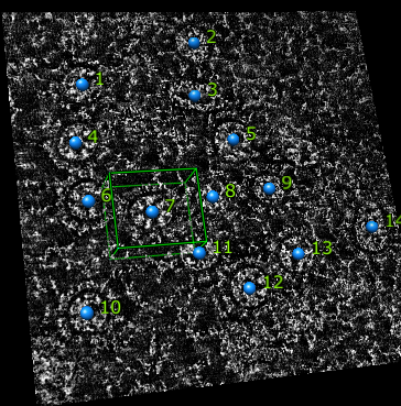
Single plane of tomogram with some virus particles marked using volume tracer.
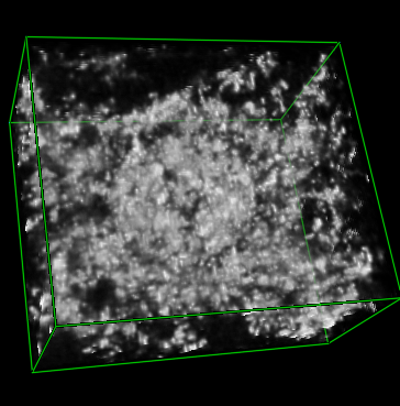
Box containing particle 7 extracted with volume dialog subregion selection. Saved to separate file for subsequent analysis.
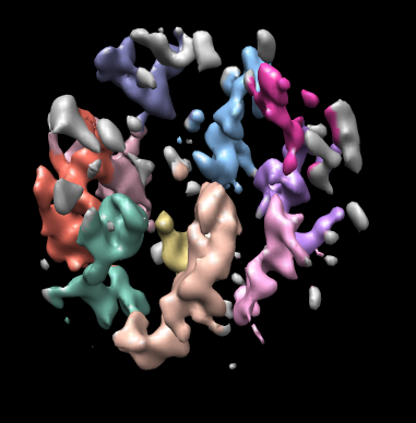
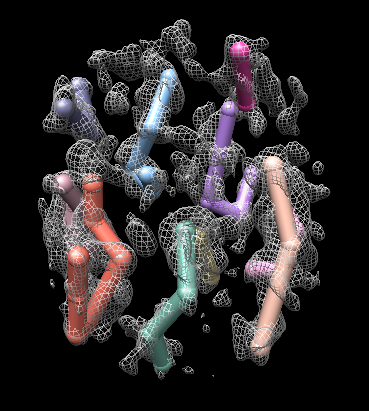
Bar-like structures in virus colored. Procedure for getting this described in following images. Used color zone dialog and bondzone command.
Movie showing bar-like structures and hand-traced core envelope. Movie made with "movie" command.
Movie showing z planes of virus particle 7.
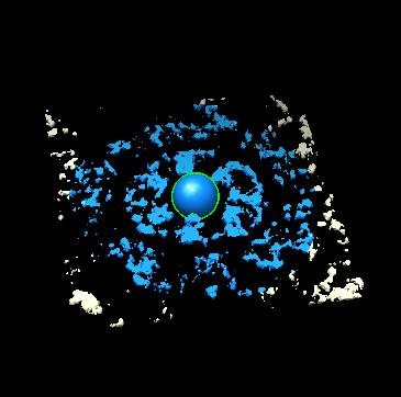
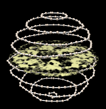
Marker placed in center of virus with volume tracer. A few data planes shown as contour surface. Color zone used color within specified radius of marker. Virus core and lipid bilayer colored blue.
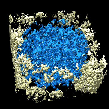
Full particle contour surface with sphere colored blue.
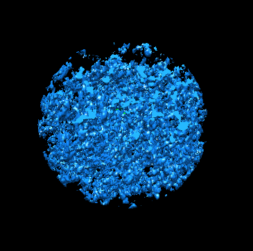
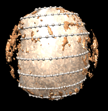
Split map button on color zone dialog used to separate blue from non-blue region.
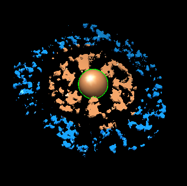
Change center marker color to brown, use color zone to separate core particle from lipid envelope.
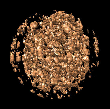
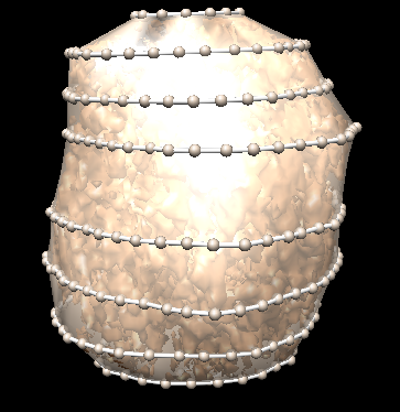
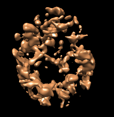
Split map used, and core shown using contour surface. Note core is not spherical so fringes on left and right in image are from lipid layer.
Hand traced loops in 9 planes to more accurately match core particle shape. Used volume tracer to hand draw loops.
Created surface from loops using Surface panel of volume tracer dialog.
Used "mask" command to extract volume within hand-traced surface.
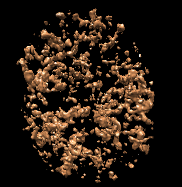
Contour surface of core particle at higher contouring level.
Used Gaussian filter (width 10, voxel size of map is set to 2, maybe nanometers) to smooth noisy core particle map.
Hand-traced bars of density using volume tracer tool.
Movie rotating view of Gaussian smoothed core data with hand-traced bars and hand-traced core particle surface.