![]() flotillin-surfs-portrait.cxs |
Flotillin Flowerpot
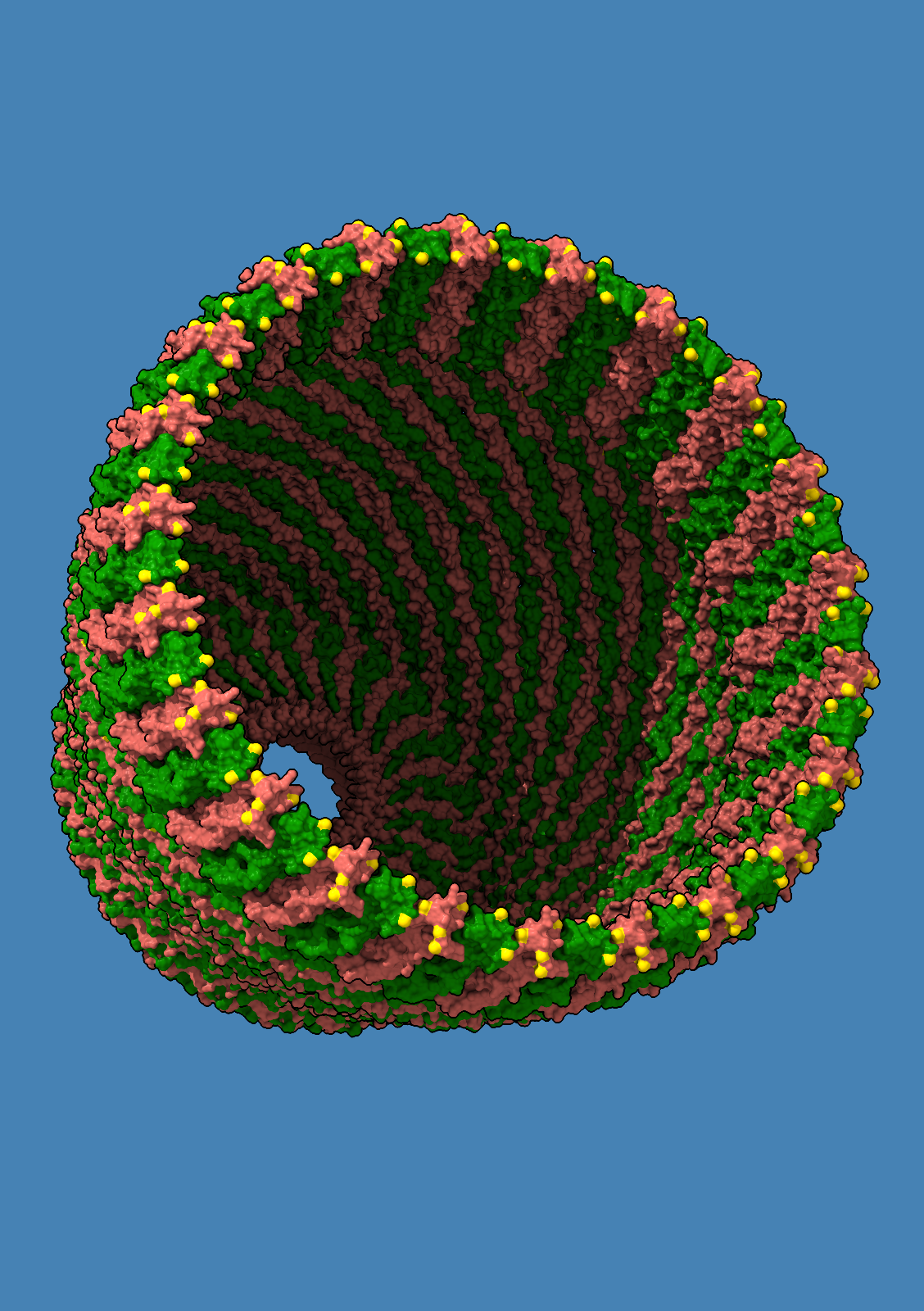
The flotillin complex is involved in membrane trafficking and the formation of membrane microdomains in mammalian cells. It has a tapered barrel or flowerpot shape formed by alternating flotillin-1 (here in green) and flotillin-2 (red) subunits, where both the wide and narrow ends may form contacts with the lipid membranes. The flowerpot rim is “decorated” with cysteine sulfur atoms (yellow). This structure is human flotillin from PDB entry 9BQ2 (Fu and McKinnon, Proc Natl Acad Sci 121:e2409334121 (2024)). [camera ortho, sym assembly 1 copies false, bg steel blue, /A indianred, /B green, gold :cys&S with atomrad 2.5, full lighting except depthcue off] |
![]() glp1-actcomp-membrane2.cxs |
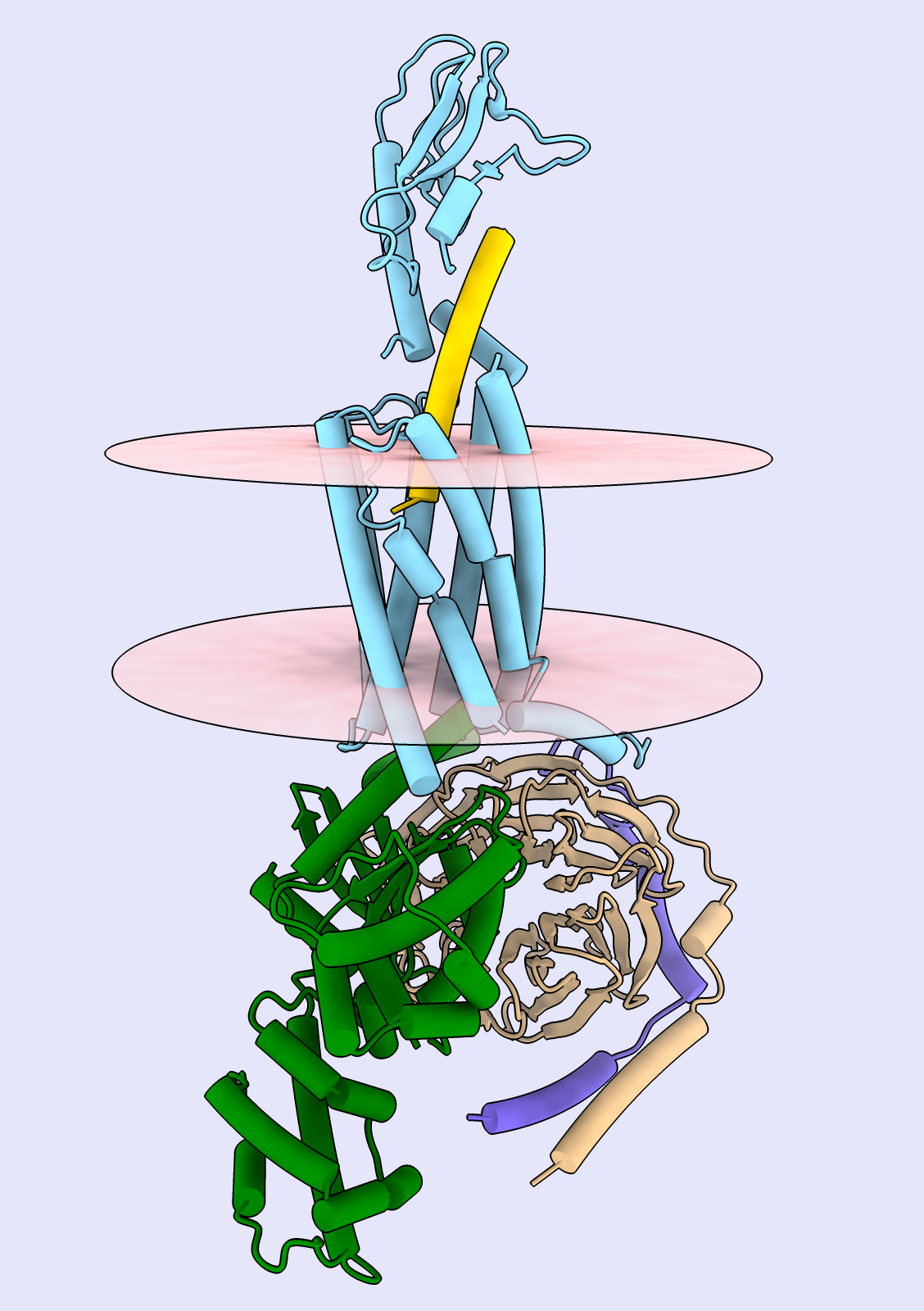
GLP-1 Signaling Complex
Human glucagon-like peptide-1 (GLP-1, yellow) is shown bound to its membrane receptor (light blue) and the associated G-protein signaling complex (α subunit green, β beige, γ purple). GLP-1 analogs such as semaglutide are widely used to treat type 2 diabetes and obesity. The structure is Protein Data Bank entry 6X18 (Zhang et al., Mol Cell 80:485 (2020)), and the transparent pink disks indicate cell membrane boundaries as estimated by the Orientations of Proteins in Membranes database (Lomize et al., Nucleic Acids Res 40:D370 (2012)). Image created with UCSF ChimeraX. [GLP-1 receptor agonists are the currently hot drugs for type 2 diabetes and obesity: Wegovy, Ozempic, etc.... so it is a very timely structure, even though the structure-determination paper is a few years old. I chose the structure with the natural ligand because I didn't want to promote any specific company or its product. *I've hidden chain N, a llama nanobody bound to the G protein; the displayed chains are all human proteins.] |
![]() origami-2views.cxs |
DNA Origami Nanopore
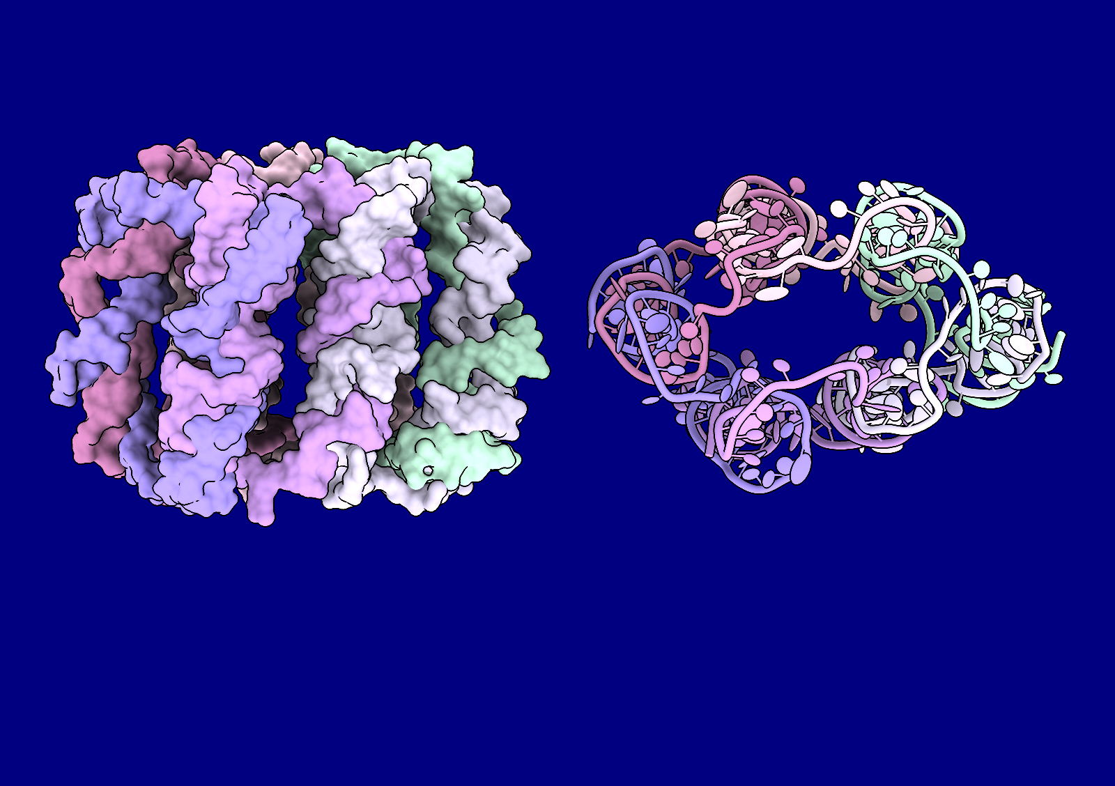
DNA origami is the art of designing sets of DNA sequences to base-pair in a specific way to create complex 3D shapes. The image shows two views of a designed six-helix bundle, where each of six unique sequences (each with its own color) base-pairs with its two neighbors. The surface view (left) shows the interlocking nature of the design. In this structure from cryo-electron microscopy (PDB 7YWL, Ahmad et al., Nat Commun 14:3630 (2023)), the bundle is somewhat squashed (right, end-on view of cartoon with ellipsoid sidechains), but it may form a more symmetrical nanopore when inserted into a membrane. |
![]() encapsulin-rdylgn.cxs |
![]() encapsulin-reverse-spectral.cxs |
Biosynthesis Nanocompartment
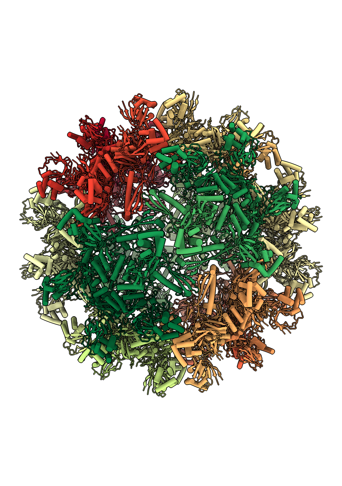
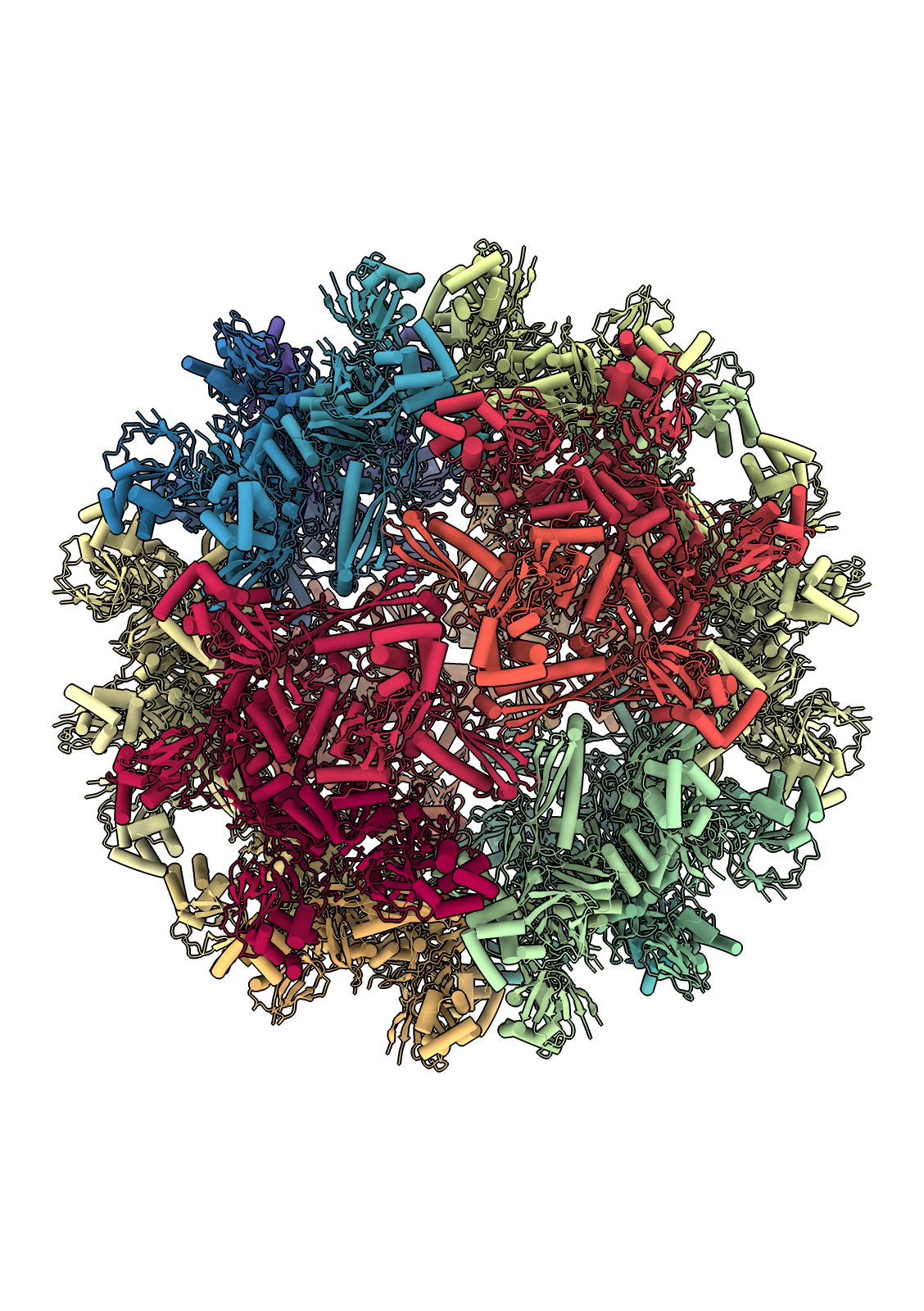
[...hmm, although festive, images look a little like the 2020 card ferritin even though the protein is totally different...] Streptomyces griseus encapsulin forms an icosahedral protein shell that encloses another protein, 2-methylisoborneol (2-MIB) synthase. 2-MIB is generated by many soil-dwelling microorganisms and may serve to repel bacterial predators or to recruit insects for spore dispersal. This structure of the (empty) encapsulin shell is PDB 9BHV (Andreas and Giessen, Nat Commun 15:9715 (2024)) . [sym #1 assembly 1 copies true, preset cylinders, light soft, graphics silhouettes true, hide pseudobonds, rainbow #2 structures palette rdylgn-11 -OR- rainbow #2 structures palette ^spectral-11] |