
|
|
|
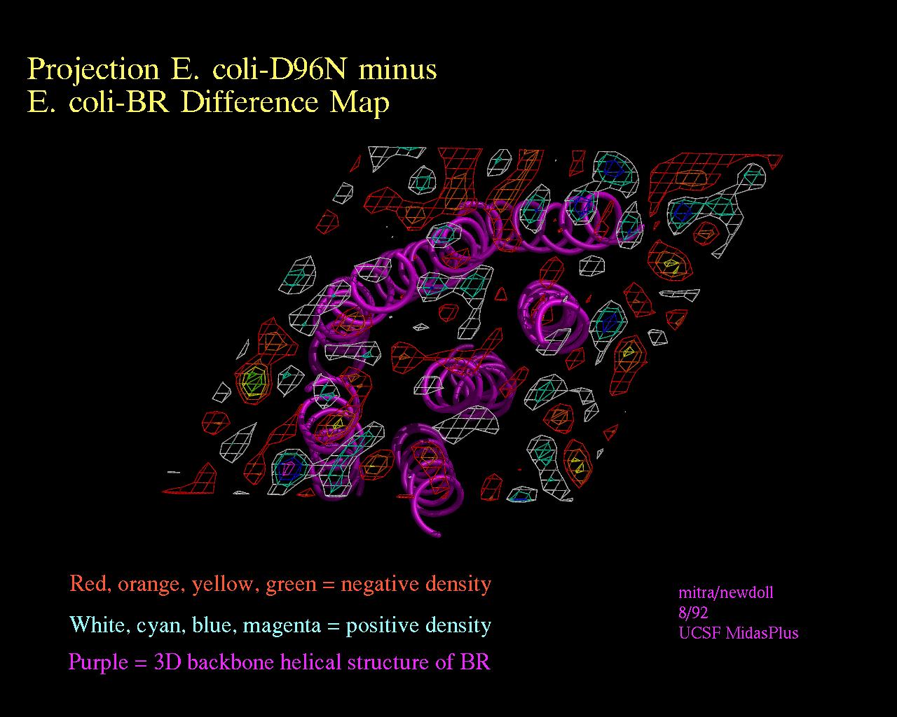
Density and helices
 JPEG
version (152KB), TIFF
version (403KB)
JPEG
version (152KB), TIFF
version (403KB)
This image is of a model of E. coli Bacteriorhodopsin superimposed with a density map obtained via x-ray diffraction.
The different colors for the density were achieved by opening several contour levels, each as a separate model number in MidasPlus. These contour levels can be displayed using the "density" delegate in MidasPlus, from XPLOR maps.
The helix was created by using neon, and the entire image was also rendered using neon. (Unless you have a fast processor, the image rendering may take a long time.) Ribbonjr is probably better suited for this type of rendering, and could also have been used to make this image. ©2004 The Regents, University of California; all rights reserved.