
Chimera Workshop
November 17-18, 2005
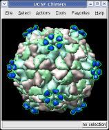
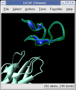
We'll look at one of the viruses that cause the common cold, Human Rhinovirus 2.
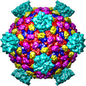
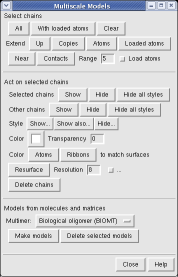
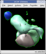
Open the PDB model 1v9u and then create 60 copies of this asymmetric unit to show a full virus capsid.
Each of the 180 capsid proteins and 60 receptor fragments is shown as a separate low resolution surface.
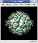
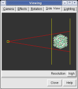
The far portion of the capsid is invisible because Chimera's near and far clip planes were positioned for the 1v9u asymmetric unit, not the full capsid. Adjust the clip planes.
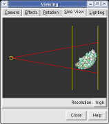
The two yellow lines in the Side View dialog are near and far clipping planes and the yellow square represents your eye position in this view from the side. Everything in front of the near clip plane or behind the far clip plane is invisible in the main Chimera window.
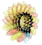
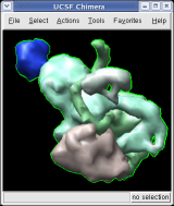
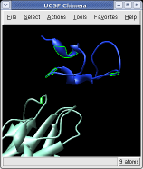
This structure shows part of the receptor found on the surface of cells in your nose that the virus recognizes. These are the chains that appear in rings of 5 near the 5-fold symmetry axes. We'll color the receptor so it stands out.

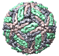
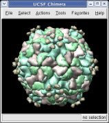
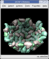
We'll hide the receptor fragment, and then hide half of the virus capsid to see the inside of the capsid.
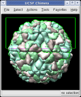
Chimera makes objects that are further from view dimmer. Adjusting the clip planes by moving the vertical yellow lines in the Side View dialog changes the distance at which the dimming effect starts and ends.
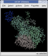
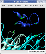
The surfaces representing each protein can be adjusted to show more detail. We will look at a single copy of the asymmetric unit, 1/60th of the icosahedral capsid.
Atomic coordinates have only been loaded for one copy of the asymmetric unit. The first 3 steps show just that copy. Some of the small blobs visible at the original surface resolution are parts of larger proteins. At the 4 angstrom resolution you can see that the green protein (VP3) has two arms that wrap around the light blue protein (VP1).
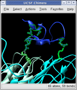
Selected proteins can be shown using atom level or residue level representations instead of surfaces.
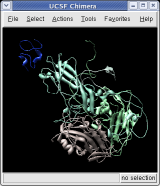
Well look specifically at the residues of the virus capsid that contact one copy of the cellular receptor fragment.
These steps find all residues within 3 angstroms of the receptor. Some contacts are with an unshown protein. To show those:
Only the atoms within 3 angstroms of the receptor are selected. To show the full residues that contain those atoms:
Note that 3 of the selected receptor residues are not really near the capsid, but instead surround a calcium ion. We should have selected both the receptor fragment and this ion in the initial contacts calculation to avoid picking up these residues.
There are about 200 virus capsid structures known at atomic resolution (ca. 2005). Below are some of the Protein Data Bank identifiers. The Virus Particle Explorer web site contains information about these virus structures.
1a34 1a6c 1al0 1al2 1aq3 1aq4 1ar6 1ar7 1ar8 1ar9 1asj 1auy 1aym 1ayn 1b35 1bbt 1bev 1bms 1bmv 1c8d 1c8e 1c8f 1c8g 1c8h 1c8m 1c8n 1cd3 1cov 1cwp 1d3e 1d3i 1d4m 1ddl 1dgi 1dnv 1dwn 1dyl 1dzl 1dzs 1e57 1e6t 1e7x 1eah 1ej6 1ev1 1f15 1f2n 1f8v 1fmd 1fod 1fpn 1fpv 1fr5 1frs 1gav 1gff 1gkv 1gkw 1gw7 1gw8 1h8j 1h8t 1hb5 1hb7 1hb9 1hdw 1he0 1he6 1hri 1hrv 1hxs 1if0 1ihm 1ijs 1jew 1js9 1k3v 1k4r 1k5m 1kuo 1kvp 1laj 1ld4 1lp3 1m06 1m0f 1m11 1m1c 1m4x 1mec 1mqt 1mst 1mva 1mvb 1mvm 1n6g 1na1 1na4 1ncq 1ncr 1nd2 1nd3 1ng0 1nn8 1nov 1ny7 1ohf 1ohg 1oop 1opo 1p58 1p5w 1p5y 1pgl 1pgw 1piv 1po1 1po2 1pov 1pvc 1qbe 1qgc 1qgt 1qju 1qjx 1qjy 1qjz 1qqp 1r08 1r09 1r1a 1rb8 1rhi 1rmu 1ruc 1rud 1rue 1ruf 1rug 1ruh 1rui 1ruj 1rvf 1s58 1sid 1sie 1smv 1stm 1sva 1tge 1thd 1tme 1tmf 1tnv 1u1y 1uf2 1upn 1v9u 1vak 1vb2 1vb4 1vba 1vbb 1vbc 1vbd 1vbe 1vrh 1w39 1wcd 1wce 1x9p 1x9t 1yc6 1zdh 1zdi 1zdj 1zdk 2bbv 2bpa 2btv 2cas 2hwb 2hwc 2hwd 2hwe 2hwf 2mev 2ms2 2plv 2r04 2r06 2r07 2rm2 2rmu 2rr1 2rs1 2rs3 2rs5 2stv 2tbv 4dpv 4rhv 4sbv 5msf 6msf 7msf
|
|
|
|
|
| Reovirus core, 1ej6 | Dengue virus, 1k4r | Ribgrass mosaic virus (helical), 1rmv | Sindbis virus, 1ld4 |
About half of the virus models from the Protein Data Bank (primarily older ones) do not contain the symmetry matrices needed to generate the capsid. When you press the Make models button you just get surfaces for the asymmetric unit. For these viruses you can download alternate coordinates from the Virus Particle Explorer web site instead of from the Protein Data Bank. Then set the Multimer type to Icosahedral symmetry, ViPER Z(2)35X(2), before pressing Make models.
Chimera
Other Sites