 JPEG version (85KB),
TIFF version (331KB)
JPEG version (85KB),
TIFF version (331KB)
JPEG version (85KB),
TIFF version (331KB)
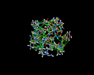
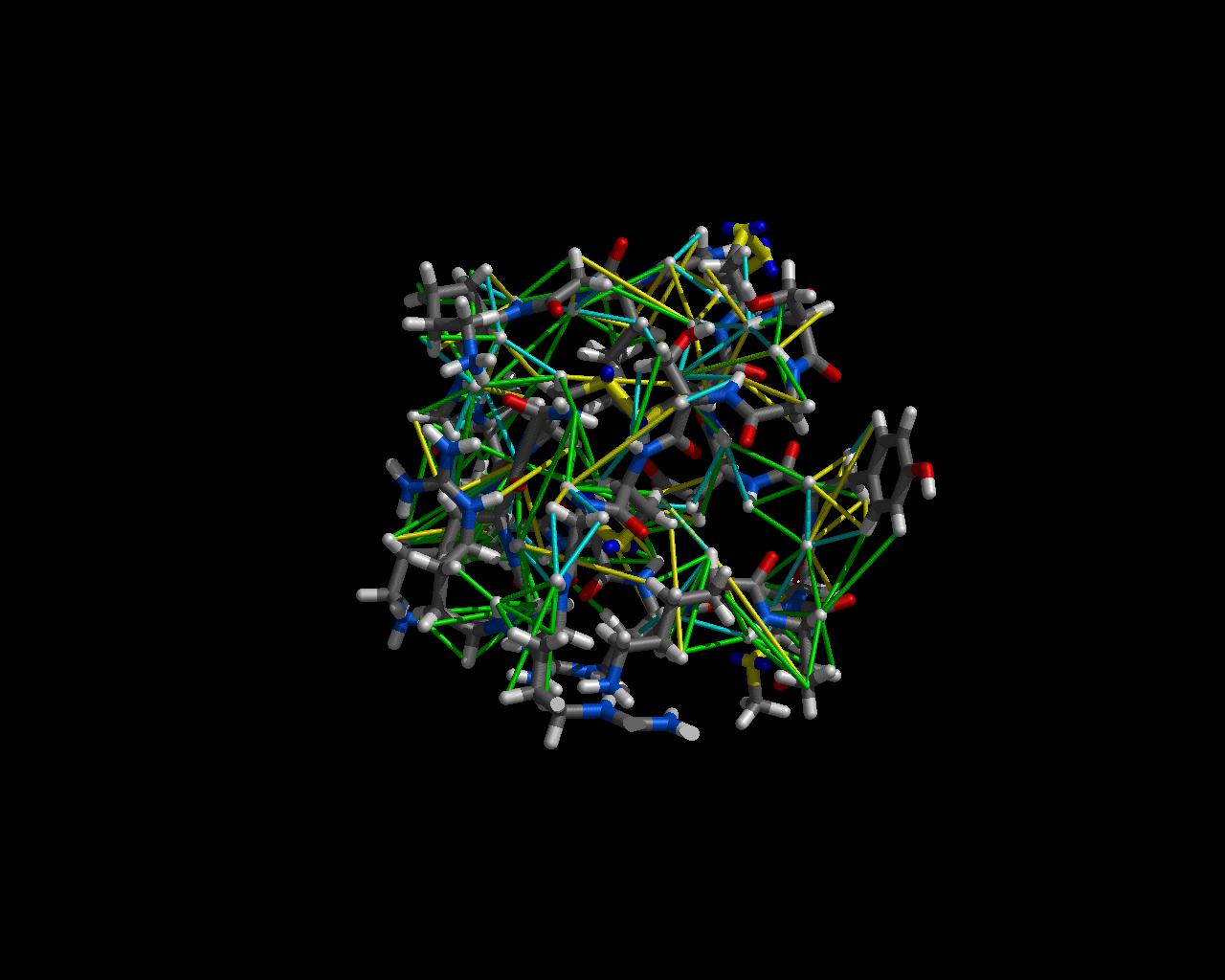
This image shows the structure of omega-conotoxin MVIIC, a small peptide found in the venom of the sea-snail Conus magus, along with the NMR constraints used to determine that structure. The structure is color-coded by atom type (via the color byatom command in MidasPlus) and the constraints (thinner tubes) are colored in the following schema:
This structure has been heavily refined and no constraint is badly violated. Bad violations would have been indicated by red (upper bound) and blue (lower bound) colored constraints.
The image was created with the MidasPlus neon utility by Dr. Shauna Farr-Jones, working in the UCSF NMR group. Dr. Farr-Jones also did the structure determination and has published a paper describing the work in the Journal of Molecular Biology.