 JPEG version (85KB),
TIFF version (1,547KB)
JPEG version (85KB),
TIFF version (1,547KB)
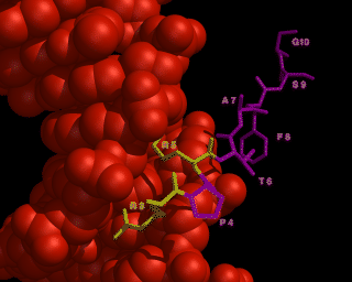
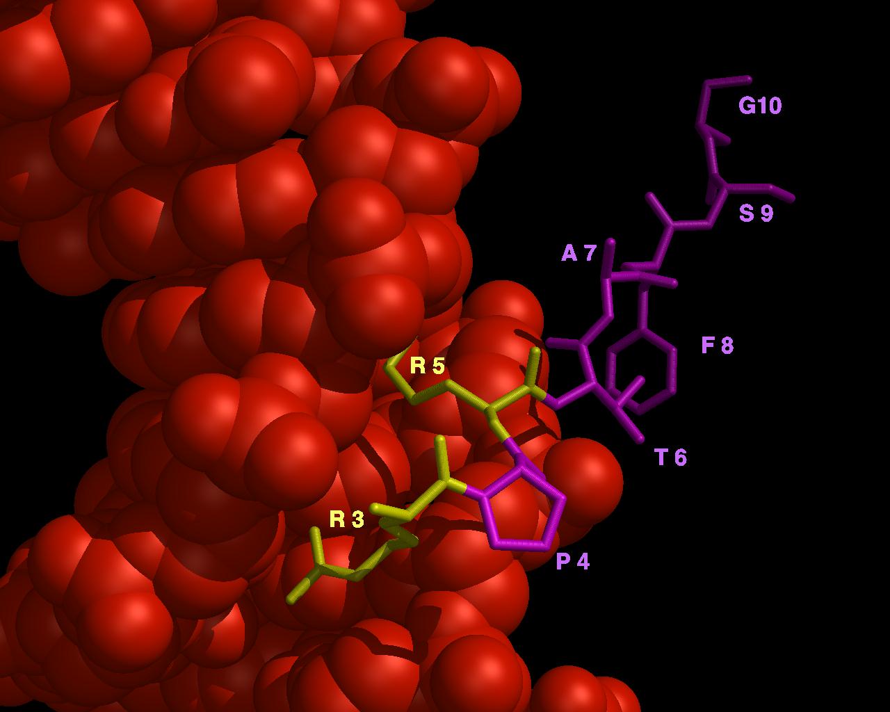
This image shows a section of protein bound to DNA.
In order to get both full sized atoms and stick representations into
the same image, an intermediate file had to be created.
- The orientation desired was chosen in MidasPlus.
- Only the DNA was displayed, the orange spheres. These coordinates
were put into a file using the "pdbrun"command.
command: pdbrun cat > image.file
- Then, only display the portion of the coordinates that are desired to
be represented as sticks (the yellow and magenta sticks).
Use the "preneon" command to put coordinates for creating sticks
into a file. These "sticks" are actually hundreds of spheres
which the MidasPlus delegate Neon determines in order to create a
cylinder between each atomic coordinate.
command: preneon cat >> image.file
DON'T FORGET TWO ">" SYMBOLS.
This is so that the "stick" coordinates will be appended
to the image.file you already created,
and not completely overwrite the file.
- Finally,
the MidasPlus delegate program Conic can be used to display the final image.
This program must now be run directly on the file, not through MidasPlus.
Get out of Midas plus. Then from any one of your windows, give the command:
conic image.file
If you want the file to be saved into an image file, give a command such as:
conic -o final.rgb image.file
See the Conic manual page for more details on saving output files,
etc.
Credits
Image by Julie Newdoll and Tom Kornberg.
Published in the Journal of Biological Chemistry, Issue of Dec 25,
pages 26813-26816, 1993, Vol 268, #36,
"Understanding the Homeodomain", Thomas B. Kornberg.
gregc@cgl.ucsf.edu / Conic and Neon / August 1994