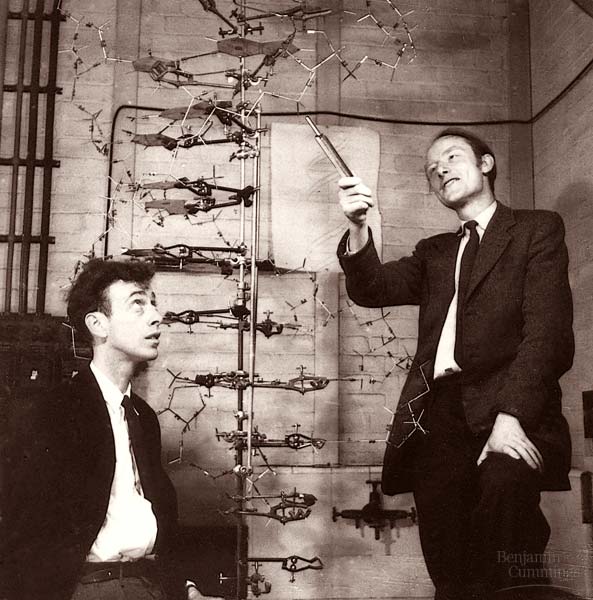
Watson & Crick model made from metal rods, 1953.
Tom Goddard
June 9, 2010
Demo for Celsius and Beyond summer camp
|
|
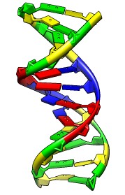
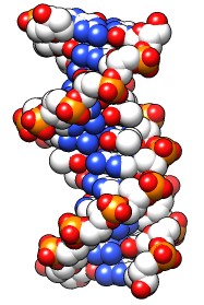
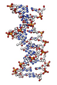
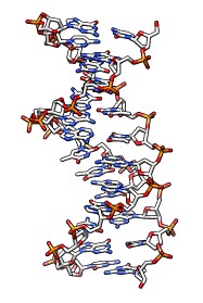
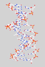
No microscope is powerful enough to see the DNA helix, so we look at models.
Watson & Crick model made from metal rods, 1953. |
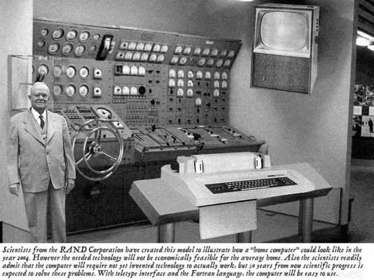
| A dream "home" computer imagined in 1950 when DNA structure solved. Molecular visualization was 20 years in the future. |
|
Today molecule models are usually viewed on computers.
|
|
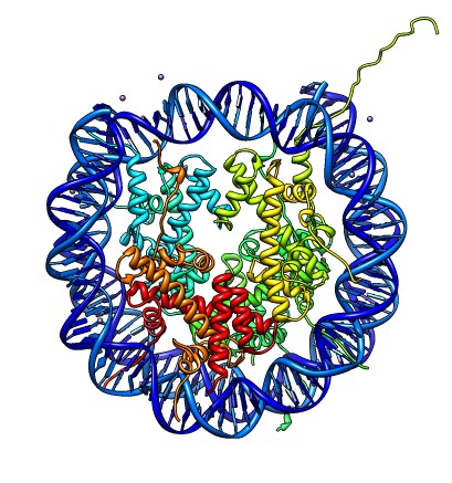
DNA in cells is wrapped around a spool (PDB 1AOI) called a nucleosome.
|
|
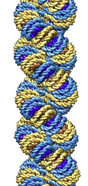
DNA spools are stacked to make filaments.
|
|
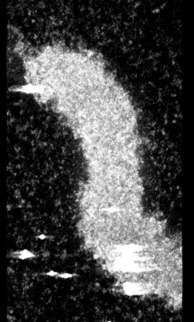
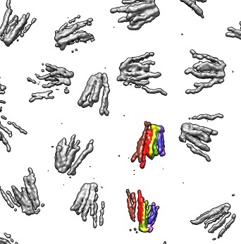
Electron microscopy -- fly chromosome tomography.
|
|
Dividing cells seen with light microscopy.
|
Show virus paper strip RNA, explain human DNA paper strip would fill entire room tightly packed.