New electron microscopy sectioning techniques can image large blocks of brain tissue.
Long range goal is to determine neural connection network for an entire fly, rat, or human brain.
Data sets will be large. Drosophila brain at 10nm voxel size = ~100 tera-bytes.
Segmentation of neurons and other structures have to be automated. The current method of hand-tracing structures plane by plane is too time consuming for large data sets. The largest neural network analyzed this way traced 300 neural cells in C. elegans taking 15 years (1980s).
Typical sample thickness for transmission electron microscopy is 0.2 microns. Thicker samples cause multiple scattering of the electron beam preventing 3-dimensional image reconstruction.
A technique introduced in 2004, serial block-face scanning electron microscopy, images thick samples by scattering the electron off the sample surface. The beam does not pass through the sample. A ~50 nm thick layer is shaved off and the next layer is imaged. The shaving is automated with a microtome installed with in the microscope high vacuum. Resolutions of 10-25 nm in x and y, and 25-50 nm in z are a bit lower than needed to resolve large molecular complexes such as ribosomes.
Serial block-face scanning electron microscopy to reconstruct three-dimensional tissue nanostructure.
Denk W, Horstmann H.
PLoS Biol. 2004 Nov;2(11):e329. Epub 2004 Oct 19.
Towards neural circuit reconstruction with volume electron microscopy techniques.
Briggman KL, Denk W.
Curr Opin Neurobiol. 2006 Oct;16(5):562-70. Epub 2006 Sep 8. Review.
There are competing experimental techniques for EM imaging of large samples.
Knife-edge scanning microscopy for imaging and reconstruction of three-dimensional anatomical structures of the mouse brain.
Mayerich D, Abbott L, McCormick B.
J Microsc. 2008 Jul;231(Pt 1):134-43.
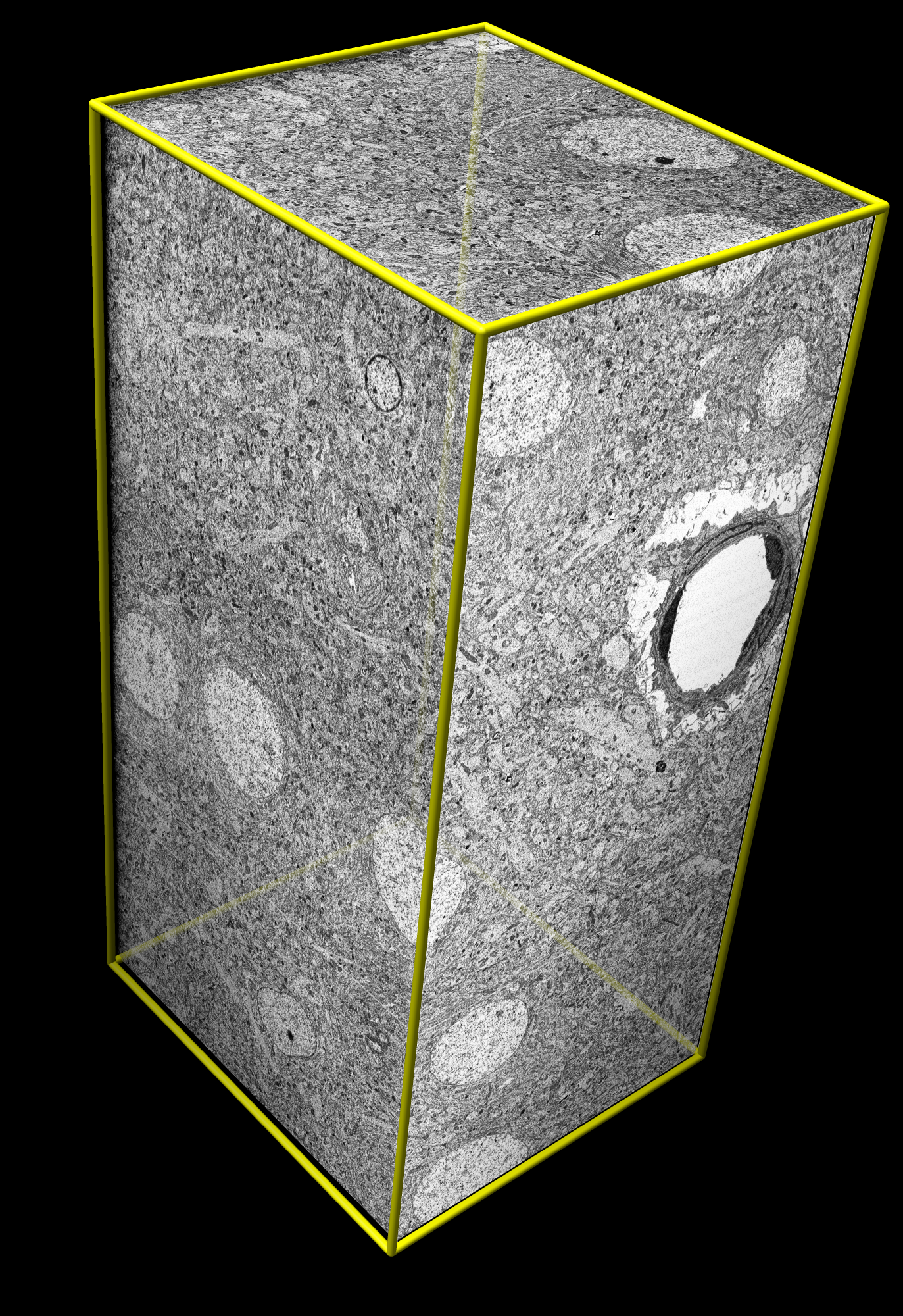
Rat cortex from Denk (2004)
Sample size 50 x 50 x 100 um.
Data size 8 Gbytes, 2048³ volume.
Voxel size 25 x 25 x 50 nm.
One voxel fits ~2 ribosomes.
Quicktime movie (25 Mb).
AVI movie (MSMPEG-4v2 encoding, 25 Mb).
