|
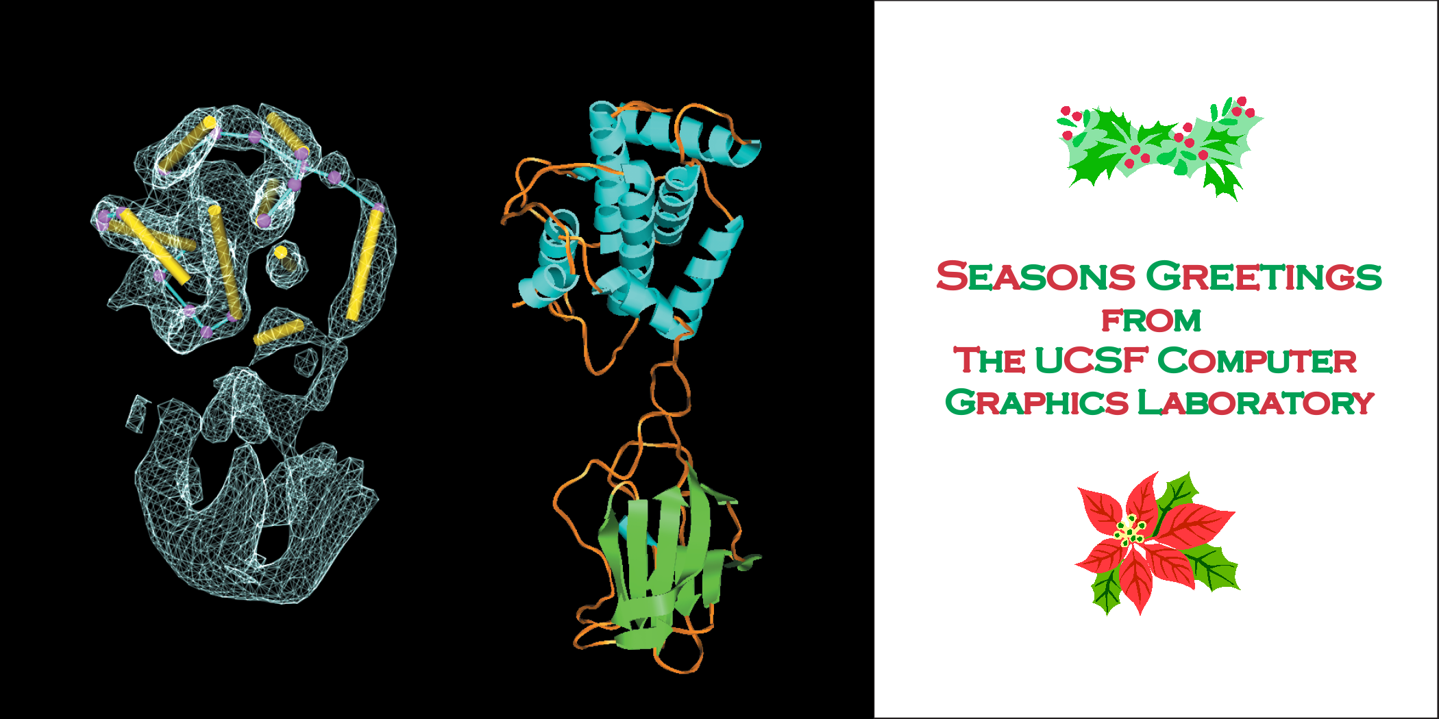
The image shows a 6.8 Å density map of rice dwarf virus P8
capsid protein beside a crystal structure of distantly related
bluetongue virus P7 capsid protein. The density map comes from
analysis of 3261 virus particles imaged by electron cryo-microscopy.
Cylinders fit to the density map indicate alpha-helix positions, and
some hand-traced connecting turns are shown. The rice dwarf virus
analysis was done at the National Center for Macromolecular Imaging
and is described in Nature Struct. Biol. 8:868-73 (2001).
The image was made with the Chimera molecular graphics program developed by the
Resource for Biocomputing, Visualization and Informatics with support
from NIH grant P41-RR01081.
© 2001 Regents of the University of California.
|